
Abstract
Peripheral nerve and nerve root involvement are rare manifestations of multiple sclerosis (MS) and may pose diagnostic challenges by mimicking other inflammatory or systemic disorders. We report the case of a 26-year-old man who presented with acute right-sided facial numbness, mild hearing impairment, and imbalance. Brain magnetic resonance imaging (MRI) demonstrated demyelinating plaques in the pons and the periventricular and subcortical white matter, accompanied by contrast enhancement of the cisternal and intrapontine segments of the right trigeminal nerve. Spinal MRI revealed focal cervical and thoracic cord lesions with symmetric enhancement of the proximal cauda equina nerve roots. Cerebrospinal fluid analysis showed a normal cell count, an immunoglobulin G index of 0.68, and a type 3 oligoclonal band pattern. The patient demonstrated marked clinical and radiologic improvement following treatment with high-dose intravenous methylprednisolone and was subsequently started on disease-modifying therapy. This case highlights a rare radiologic manifestation of MS and underscores the importance of considering MS in the differential diagnosis of concurrent cranial nerve and cauda equina root enhancement.
Keywords: Multiple sclerosis, trigeminal nerve, cauda equina, cranial nerve enhancement, magnetic resonance imaging
Introduction
Multiple sclerosis (MS) is a chronic immune-mediated demyelinating disease of the central nervous system (CNS). Magnetic resonance imaging (MRI) plays a crucial role not only in establishing the diagnosis but also in excluding imaging mimics, particularly when atypical findings are encountered (). Trigeminal nerve involvement is uncommon and may occur with or without classic trigeminal neuralgia (, ). Spinal cord involvement in MS typically presents as short-segment lesions, which may help distinguish MS from other demyelinating disorders (). Cranial nerve enhancement has been increasingly recognized with the use of high-resolution MRI, and a pattern-based approach can aid in differentiating demyelinating involvement from infectious, neoplastic, or granulomatous etiologies (). In contrast, cauda equina nerve root enhancement is exceedingly rare in MS and more commonly suggests infectious, granulomatous, neoplastic, or systemic autoimmune conditions (). We present a rare case of MS with concurrent trigeminal nerve and proximal cauda equina root enhancement and discuss the key differential diagnostic considerations.
Case Report
Written informed consent was obtained from the patient for publication of this case report and the accompanying images.
A 26-year-old man presented with a 1-week history of right-sided facial numbness accompanied by mild right-sided hearing impairment, facial heaviness, imbalance, and fatigue. Neurologic examination revealed hypoesthesia in the distribution of the right trigeminal nerve, with no other focal neurologic deficits.
Post-contrast MRI demonstrated linear enhancement involving the cisternal and intrapontine segments of the right trigeminal nerve ( and ). Brain MRI revealed multiple demyelinating plaques in the periventricular, subcortical, and pontine regions ( and ). Spinal MRI demonstrated focal lesions in the cervical and thoracic spinal cord ( and ), along with symmetric enhancement of the proximal cauda equina nerve roots ( and ).
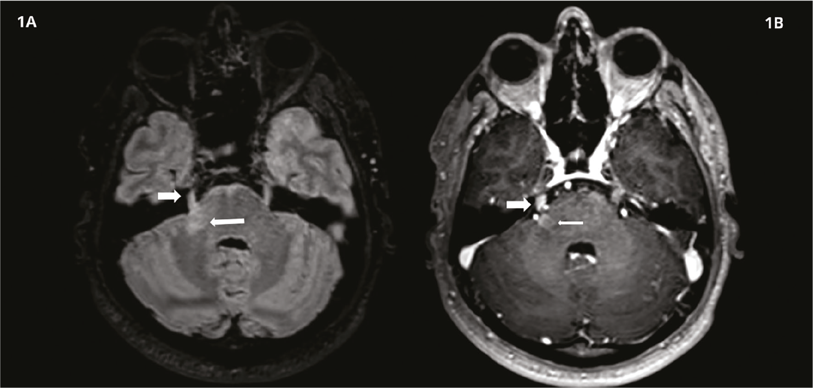
(A) Axial FLAIR image showing thickening and edema of the cisternal segment of the right trigeminal nerve (thick arrow) and an active demyelinating plaque along the intrapontine pathway (thin arrow). (B) Post-contrast T1-weighted image demonstrating enhancement of both the cisternal segment of the trigeminal nerve (thick arrow) and the demyelinating plaque (thin arrow). Subpial plaques along the anterior pons show no contrast enhancement
MRI: Magnetic resonance imaging
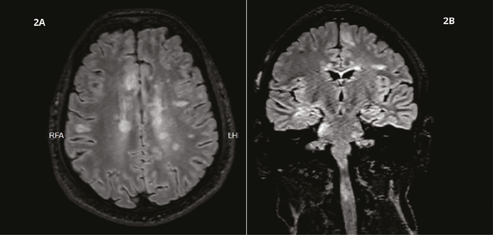
(A) Axial FLAIR image showing multiple demyelinating plaques in the periventricular, centrum semiovale, and subcortical regions. (B) Coronal FLAIR image demonstrating demyelinating plaques involving the cerebral parenchyma, brainstem, and cervical spinal cord
FLAIR: Fluid-attenuated inversion recovery
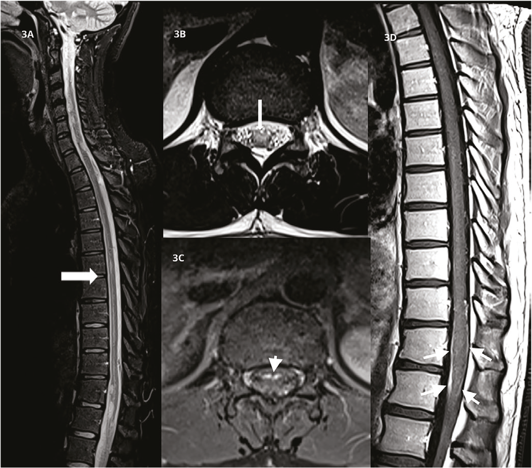
(A) Sagittal STIR image showing extensive demyelinating plaques and edema (arrow) along the anterior subpial surface of the conus medullaris. (B) Axial T2-weighted turbo spin-echo image showing a conus medullaris plaque (arrow) localized to the anterior half of the spinal cord. (C,D) Post-contrast axial and sagittal images demonstrating symmetric enhancement of the proximal cauda equina nerve roots (arrows)
STIR: Short-tau inversion-recovery
Cerebrospinal fluid analysis revealed a normal cell count without pleocytosis, an immunoglobulin G (IgG) index of 0.68, and a type 3 oligoclonal band pattern characterized by identical oligoclonal IgG bands in both serum and cerebrospinal fluid, together with additional cerebrospinal fluid-restricted bands. Serum angiotensin-converting enzyme levels were within normal limits. Antinuclear antibody testing showed weak positivity (1:100), whereas anti-double-stranded DNA antibodies, the extractable nuclear antigen panel, anti-aquaporin-4 antibodies, and anti-myelin oligodendrocyte glycoprotein antibodies were negative. Additional laboratory investigations performed to exclude infectious, systemic inflammatory, and autoimmune etiologies were unremarkable.
The patient was treated with intravenous methylprednisolone (1 g/day for 7 days), resulting in near-complete clinical recovery. Follow-up MRI demonstrated resolution of the trigeminal nerve and cauda equina root enhancement. Disease-modifying therapy with ocrelizumab was subsequently initiated.
Discussion
Cranial nerve enhancement in MS is uncommon but is being increasingly recognized with the widespread use of high-resolution MRI. Trigeminal nerve involvement most often reflects inflammatory demyelination at the root entry zone or cisternal segment and may occur even in the absence of trigeminal neuralgia (, ). A pattern-based approach to cranial nerve enhancement is essential for distinguishing demyelinating involvement from infectious, neoplastic, and granulomatous etiologies ().
Spinal cord lesions in MS are typically short-segment and peripherally located, most commonly involving the cervical and thoracic spinal cord. This characteristic distribution can aid in differentiating MS from other antibody-mediated demyelinating disorders (). Although cauda equina root enhancement is rare in MS, its coexistence with cranial nerve enhancement necessitates careful exclusion of alternative diagnoses, including connective tissue diseases and overlap syndromes (, ).
Although MS is classically defined as a demyelinating disease of the CNS, pathologic and imaging studies suggest that inflammatory activity may extend beyond conventional CNS boundaries, particularly at sites where central and peripheral myelin interface. These transitional regions, such as the root entry zones of cranial and spinal nerves, may be especially vulnerable to inflammatory infiltration and demyelination. In addition, perivascular inflammation, blood-brain barrier disruption, and immune-mediated processes involving both adaptive and innate immune responses have been implicated in lesion formation and extension in MS. Collectively, these mechanisms may provide a plausible explanation for the rare occurrence of nerve root enhancement, including cauda equina involvement, in otherwise typical cases of MS ().
Previous reports have described cranial nerve enhancement in MS, most commonly involving the trigeminal nerve (), as well as occasional peripheral nerve or nerve root involvement in atypical presentations (, ). In a large MRI cohort, cranial nerve enhancement was observed in 8.2% of patients, with trigeminal nerve involvement representing the most frequent finding (). However, the simultaneous presence of both trigeminal nerve enhancement and cauda equina root involvement has rarely been documented. Compared with previously reported cases, our patient demonstrated a combination of cranial and spinal peripheral involvement in conjunction with typical demyelinating lesions of the brain and spinal cord, thereby expanding the spectrum of atypical radiologic manifestations of MS.
Trigeminal neuralgia is a recognized manifestation of MS, with a pooled prevalence of approximately 3.4% reported in a recent systematic review and meta-analysis (). Furthermore, cranial nerve enhancement has been associated with greater lesion burden and a more severe disease course, underscoring its potential clinical significance (). To our knowledge, simultaneous contrast enhancement of the trigeminal nerve and proximal cauda equina nerve roots has rarely been reported in patients with MS.
Conclusion
Concurrent trigeminal nerve and cauda equina root enhancement represents a rare manifestation of active MS. Recognition of this atypical imaging pattern is crucial to avoid misdiagnosis and to ensure timely initiation of appropriate disease-modifying therapy.
Footnotes
Ethics
Informed Consent: Written informed consent was obtained from the patient for the publication of anonymized clinical and radiologic data.
Authorship Contributions
Concept: T.C., O.A.D., Design: T.C., Data Collection or Processing: T.C., A.C., Analysis or Interpretation: T.C., A.C., Literature Search: O.A.D., Writing: T.C., O.A.D.
Conflict of Interest: No conflict of interest was declared by the authors.
Financial Disclosure: The authors declared that this study received no financial support.
References
Geraldes R, Ciccarelli O, Barkhof F, De Stefano N, Enzinger C, Filippi M, Hofer M, Paul F, Preziosa P, Rovira A, DeLuca GC, Kappos L, Yousry T, Fazekas F, Frederiksen J, Gasperini C, Sastre-Garriga J, Evangelou N, Palace J; MAGNIMS study group. The current role of MRI in differentiating multiple sclerosis from its imaging mimics. Nat Rev Neurol. 2018;14:199-213. Erratum in: Nat Rev Neurol. 2018;14:213.
Nakashima I, Fujihara K, Kimpara T, Okita N, Takase S, Itoyama Y. Linear pontine trigeminal root lesions in multiple sclerosis: clinical and magnetic resonance imaging studies in 5 cases. Arch Neurol. 2001;58:101-104.
Chaudhary K, Zekavaty S, Xu K, Mahesh S, Eppurath A, Bordia R. Involvement of cranial nerve V (trigeminal nerve) in multiple sclerosis. Int J Case Rep Images. 2023;14:134-137
Marrodan M, Gaitán MI, Correale J. Spinal cord involvement in MS and other demyelinating diseases. Biomedicines. 2020;8:130.
Moltoni G, Romano A, Blandino A, Palizzi S, Romano A, D’Arrigo B, Guarnera A, Dellepiane F, Frezza V, Gagliardo O, Tari Capone F, Grossi A, Trasimeni G, Bozzao A. Extra-axial cranial nerve enhancement: a pattern-based approach. Radiol Med. 2024;129:118-132.
Shakour RL, Tascione O, Carberry N, Flores-Gonzalez R. Exploring overlap syndromes: an atypical case of multiple sclerosis with anti-Sjogren’s syndrome type B antibody. J Med Cases. 2024;15:387-395.
Frosch M, Kremers N, Lisko K, Urbach H, Prinz M, Taschner CA. Freiburg neuropathology case conference: a 42-year-old patient with progressive neurological deficits, multiple brain lesions and accompanying affection of peripheral nerves. Clin Neuroradiol. 2021;31:529-535.
Reich DS, Lucchinetti CF, Calabresi PA. Multiple sclerosis. N Engl J Med. 2018;378:169-180.
Haider L, Chan WE, Olbert E, Mangesius S, Dal-Bianco A, Leutmezer F, Prayer D, Thurnher M. Cranial nerve enhancement in multiple sclerosis is associated with younger age at onset and more severe disease. Front Neurol. 2019;10:1085.
Houshi S, Tavallaei MJ, Barzegar M, Afshari-Safavi A, Vaheb S, Mirmosayyeb O, Shaygannejad V. Prevalence of trigeminal neuralgia in multiple sclerosis: a systematic review and meta-analysis. Mult Scler Relat Disord. 2022;57:103472.